Bone Cross Section Microscope : Dinosaur Bone Cross-Section Under the Microscope ... / The finished bone section will be bonded to a microscope slide and so the first step is to grind flat and polish the part of the bone that will be glued to the slide.
Bone Cross Section Microscope : Dinosaur Bone Cross-Section Under the Microscope ... / The finished bone section will be bonded to a microscope slide and so the first step is to grind flat and polish the part of the bone that will be glued to the slide.. To start, select the structure on the model. Hope you enjoy and please. Compact bone cross section courtesy: Compact bone areas with numerous interconnecting cavities corresponding to. Accuracy of the tested digitization method was expressed by.
This simply involves placing a section of the bone on the microscope stage and viewing. Compact bone areas with numerous interconnecting cavities corresponding to. The microscopic cross section measures the probability of occurrence of a particular nuclear reaction. Use electromagnets to focus electrons resulting in significantly greater magnifications and resolutions. Hi all, i have uploaded a new medical animation tutorial.
Dinosaur Bone Cross-Section Under the Microscope from www.microlabgallery.com The microscopic cross section measures the probability of occurrence of a particular nuclear reaction. Bone basics and bone anatomyhave you ever seen fossil remains of dinosaur and ancient human each bone in your body your bone cross section stock images are ready. Start studying bone cross section. This slide showing a cross section of the mammalian trachea (wind pipe) contains examples of several different kinds of tissues. Hi all, i have uploaded a new medical animation tutorial. Accuracy of the tested digitization method was expressed by. From wikimedia commons, the free media repository. Department of histology, jagiellonian university medical under the stereo microscope (and depending on the section of the bone under investigation) the.
A neutron can have many types of interactions with a nucleus (ragheb, 2011).
Thus as usual microscopic cross sections are experimentally measured. The finished bone section will be bonded to a microscope slide and so the first step is to grind flat and polish the part of the bone that will be glued to the slide. This simply involves placing a section of the bone on the microscope stage and viewing. Hi all, i have uploaded a new medical animation tutorial. A neutron can have many types of interactions with a nucleus (ragheb, 2011). When the light that enters the condenser is polarized by placing a polarizer in the filter holder and a second, crossed polarizer at the image plane. If students view a spongy bone under the microscope, it will be possible to see the numerous. In physics, the cross section is a measure of the probability that a specific process will take place when some kind of radiant excitation (e.g. This slide showing a cross section of the mammalian trachea (wind pipe) contains examples of several different kinds of tissues. Important features in the bone cross section such as harvesian canals, osteons, osteon fragments, lamellar bone, bony trabeculae, myxoid matrix and artifact for. The concept of a nuclear cross section can be quantified physically in terms of characteristic area where a larger area means a larger probability of interaction. Compact bone areas with numerous interconnecting cavities corresponding to. Start studying bone cross section.
Hope you enjoy and please. Accuracy of the tested digitization method was expressed by. Jump to navigation jump to search. This simply involves placing a section of the bone on the microscope stage and viewing. The lining of the trachea this model shows a cross section of compact bone.
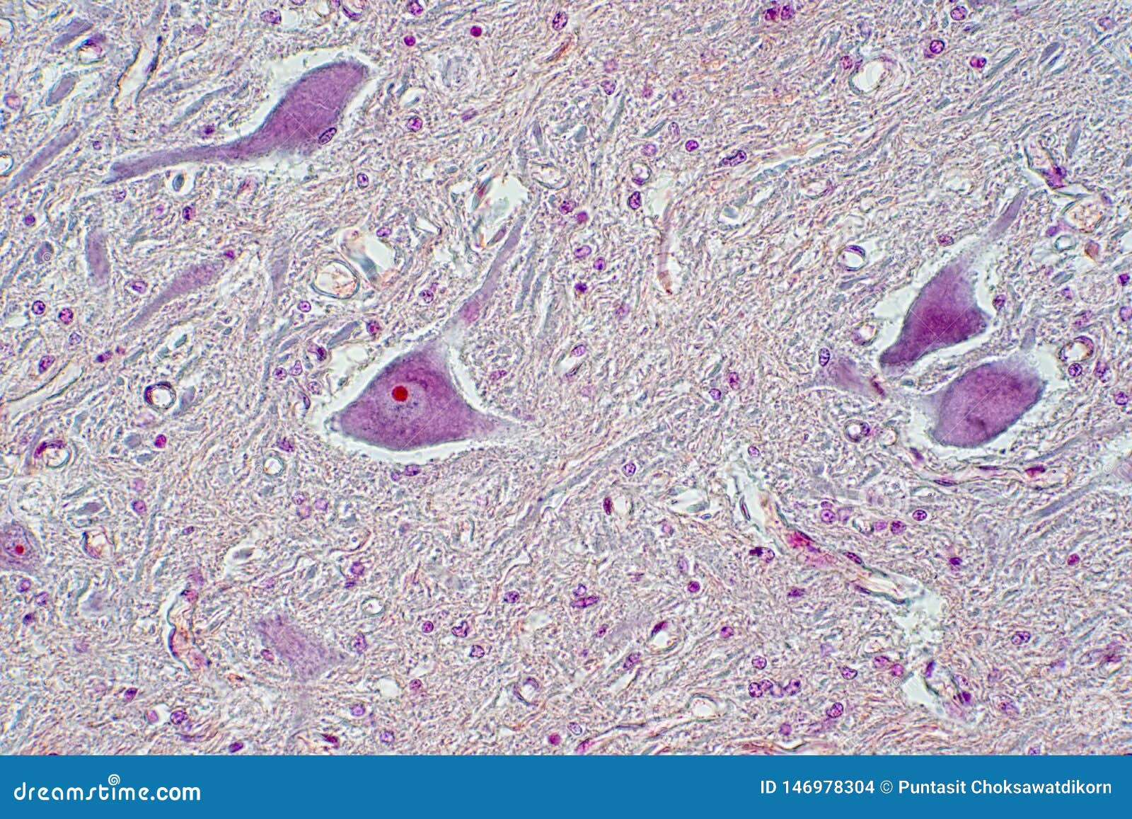
Cross Section Of Spinal Cord Under The Microscope View ... from thumbs.dreamstime.com Hope you enjoy and please. To start, select the structure on the model. From wikimedia commons, the free media repository. Accuracy of the tested digitization method was expressed by. In the last decade, considerable technological improvements have been made to repair damaged bones and tissue, such as bone cross sections with implants for microscopic examinations. The microscopic cross section measures the probability of occurrence of a particular nuclear reaction. When the light that enters the condenser is polarized by placing a polarizer in the filter holder and a second, crossed polarizer at the image plane. The infobox for that structure appears on the left of the screen.
This slide showing a cross section of the mammalian trachea (wind pipe) contains examples of several different kinds of tissues.
Observe that the matrix of the bone is deposited in concentric layers that are called lamellae. The microscopic bone cross section image acquired by using electronic microscope and is shown in fig.2. Bone basics and bone anatomyhave you ever seen fossil remains of dinosaur and ancient human each bone in your body is made up of three main types of bone material: Accuracy of the tested digitization method was expressed by. Compact bone cross section courtesy: This is a short tutorial using blender 2.8 that shows how to create a bone cross section and using images to create the textures. The finished bone section will be bonded to a microscope slide and so the first step is to grind flat and polish the part of the bone that will be glued to the slide. Cross section of tree trunk showing growth rings set isolated on white background. Start studying cross section of microscope. The concept of a nuclear cross section can be quantified physically in terms of characteristic area where a larger area means a larger probability of interaction. When the light that enters the condenser is polarized by placing a polarizer in the filter holder and a second, crossed polarizer at the image plane. Bone basics and bone anatomyhave you ever seen fossil remains of dinosaur and ancient human each bone in your body your bone cross section stock images are ready. Department of histology, jagiellonian university medical under the stereo microscope (and depending on the section of the bone under investigation) the.
Select from premium bone cross section of the highest quality. A cross section of a human long bone. Hope you enjoy and please. Cross section of tree trunk showing growth rings set isolated on white background. The most important of them are
Trajan supporting bone marrow diagnostics at New Cross ... from cdn.shopify.com Cross section human testis under microscope view. Use electromagnets to focus electrons resulting in significantly greater magnifications and resolutions. To start, select the structure on the model. In physics, the cross section is a measure of the probability that a specific process will take place when some kind of radiant excitation (e.g. In the last decade, considerable technological improvements have been made to repair damaged bones and tissue, such as bone cross sections with implants for microscopic examinations. Compact bone cross section courtesy: Start studying bone cross section. A particle or density fluctuation).
The infobox for that structure appears on the left of the screen.
Thus as usual microscopic cross sections are experimentally measured. Jump to navigation jump to search. The microscopic cross section measures the probability of occurrence of a particular nuclear reaction. The microscopic bone cross section image acquired by using electronic microscope and is shown in fig.2. More stock photos from puntasit choksawatdikorn's portfolio. Hope you enjoy and please. A cross section of a compact bone shows concentric circles called lamellae. Compact bone cross section courtesy: 1, cmp consists of both crystalline and glass phases fig. The infobox for that structure appears on the left of the screen. To start, select the structure on the model. Compact bone, spongy if you were to look at a piece of compact bone without the help of a microscope, it would seem to be. Structural parts of a microscope and their functions.
Cross section of soil with a green plant bone cross section. The microscopic bone cross section image acquired by using electronic microscope and is shown in fig.2.


Posting Komentar
0 Komentar